Find out what the differences and similarities are between these two spine conditions. One way of protecting your heart that you might not be aware of is by getting the COVID-19 vaccine.
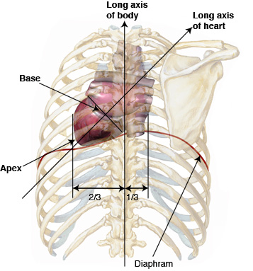
The Location Size And Shape Of The Heart
As part of the bony thorax the thoracic vertebrae help protect the internal viscera such as the heart lungs and oesophagus.

. An anterior surgical approach of the CTJ can be challenging due to. The cervical spine series is a set of radiographs taken to investigate the bony structures of the cervical spine albeit commonly replaced by the CT the cervical spine series is an essential trauma radiograph for all radiographers to understand. The thoracic spine is the second segment of the vertebral column located between the cervical and lumbar vertebrae.
Sagittal coronal and transverse slices. Posterior Tilt Hip Biomechanics. Surgery of the C7-T1 vertebral level may be performed from the front anterior or back posterior of the neck.
A Baylor College of Medicine cardiologist says that SARS-CoV-2 causes inflammation and injury that can damage a number of organs and that includes the heart. As the upper part of the pelvis is pulled backward the bottom part of the pelvis is pulled forward. A chapter on joints and ligaments of the spine including atlanto-axial joints costovertebral joints and.
Anatomy of the lumbar spine using cross-sectional imaging MR T1 and T2 weighted. Tibialis posterior is involved in movements at two different joints as follows. The sacrum and coccyx in lateral superior anterior and posterior views as well as sagittal and axial sections of the sacrum and coccyx.
Indications These views are specialized projections to provide functional tests 1 of lumbar spine instability often in the context of spondy. The lumbar spine flexion and extension views images the lumbar spine which consists of five vertebrae. Cervical spine radiographs are indicated for a variety of settings including 1-3.
The cervical portion of the spine is an important one anatomically and clinically. Inversion of the foot at the subtalar joint. It consists of twelve vertebrae which are separated by fibrocartilaginous intervertebral discs.
Proximity to vital organs such as the heart and lungs. In a posterior tilt the upper part of the pelvis is positioned behind the imaginary vertical plumb line or at least as can be the case during exercise is moving in that direction. The cervical spine also allows passage of important vasculature to reach the brain and provides attachment sites.
It is within this region that the nerves to the arms arise via the brachial plexus and where the cervical plexus forms providing innervation to the diaphragm among other structures. A posterior disc bulge is not the same as a herniated disc. Plantar flexion of the foot at the talocrural ankle joint.
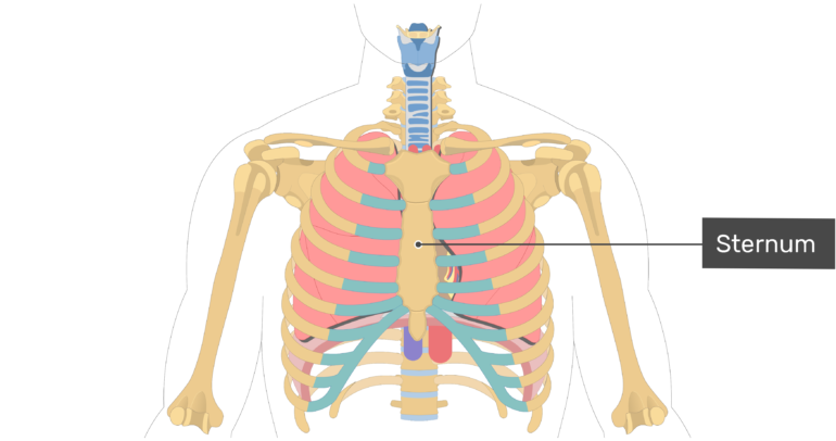
Osteology Bones and Bony Landmarks of the Axial Skeleton Bones and Bony Landmarks of the Appendicular Skeleton Musculature Musculature of the Head and Neck. Beckers Orthopedic Spine Review is a leading resource offering news and analysis on business and legal issues relating to orthopedic and spine practices. Lower visibility due to the presence of bones such as the first rib the collar bone and the breast bone.
Through its action on the ankle joint tibialis posterior helps the other more powerful foot flexors to elevate the heel when the foot is planted on the ground.
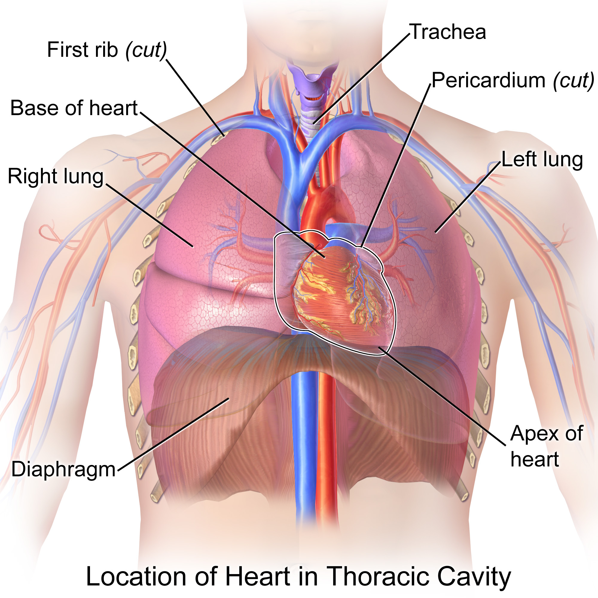
Is The Heart Located Posterior Or Medial To The Lungs Socratic
Anatomy Tutorial Posterior Atlas Of Human Cardiac Anatomy

Heart Anatomy Anatomy And Physiology Ii
Heart Anatomy Anatomy And Physiology Ii

Heart Anatomy Anatomy And Physiology Ii
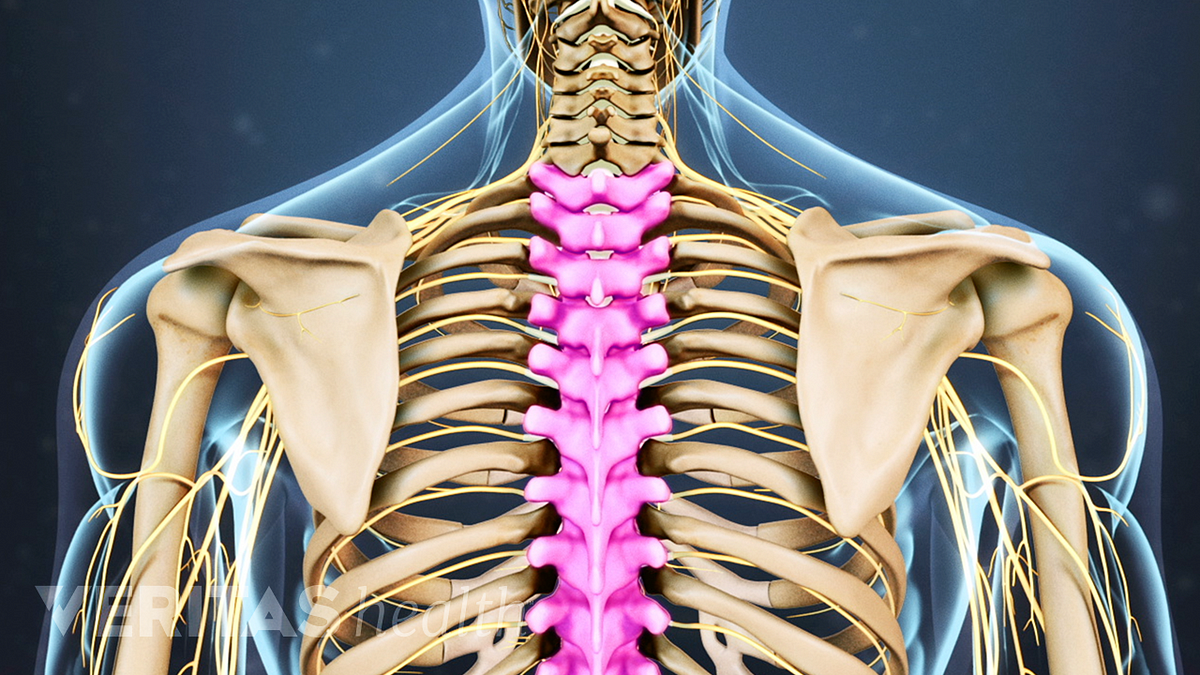
Thoracic Spine Anatomy And Upper Back Pain

Positioning Of The Heart

Posterior View Of The Organs Within The Abdominal Cavity The Lumbar Download Scientific Diagram
0 comments
Post a Comment